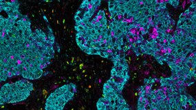
Immunofluorescence
Immunofluorescence (IF) is an immunostaining technique that combines the use of antibodies with fluorescence imaging to visualize specific proteins and other biomolecules within a tissue or cell sample. IF is a powerful technique that can be used with a variety of samples including fixed whole cells, paraffin-embedded cell pellets, and frozen or paraffin-embedded tissue samples.

What is Immunofluorescence?
IF is an immunoassay technique that uses antibodies labeled either directly or indirectly with a fluorophore to visualize proteins and other biomolecules. Though the terms IF and immunohistochemistry (IHC) are often used interchangeably, they carry distinct meanings, with IHC denoting the use of an enzymatic reaction resulting in a visible precipitate to visualize an antibody. Since its initial development, IF has grown into a powerful tool enabling the visualization and colocalization of many proteins and biomolecules within a single tissue with an accompanying array of fluorophores that cover the entire visual spectrum.
How Does Immunofluorescence Work?
IF protocols can be divided into three major steps: sample processing, staining, and imaging. Sample processing occurs prior to staining and typically involves fixing tissue samples in a cross-linking agent such as paraformaldehyde to preserve tissue morphology. Tissues are then dehydrated and embedded into paraffin blocks enabling tissues to be cut into tissue sections and affixed to a glass slide for long-term storage. When slides are ready to be stained, they are processed in a clearing agent to remove excess paraffin and are rehydrated.
Staining protocols usually start with a blocking step consisting of either a protein or serum block to reduce non-specific binding. Sections are next incubated with an antibody specific to the protein or biomolecule of interest. At this point, there are two main types of IF protocols: direct and indirect. If the primary antibody is conjugated with a fluorophore, then it is considered direct IF. An example of indirect IF is when the primary antibody is unconjugated and a targeted secondary antibody is conjugated with a fluorophore. Indirect IF has the advantage of increasing signal enabling the detection of less abundant proteins, but at the cost of increased complexity and potential background.
Once slides are finished staining, they are then imaged with an epi-fluorescent, confocal, or multispectral microscope. Depending on the number and type of fluorophores used, it may be necessary to do spectral unmixing to ensure that each marker is accurately quantitated. Although there is a plethora of fluorophores to choose from, many have overlapping emission spectra limiting their use in multiplexing. Spectral unmixing is a powerful technique that distinguishes fluorophores not only from other fluorophores being used that have overlapping spectra but even background fluorescence.
Why Use Immunofluorescence?
IF offers a powerful method for visualizing multiple proteins within a single sample. With advances in confocal microscopy and multispectral imaging, it is now possible to stain more than nine antigens in a single tissue, which is impossible with traditional IHC.
Unlike IHC, IF also allows users to understand co-expression and spatial relationships between multiple proteins and biomolecules through co-localization, while still preserving tissue context.
Immunofluorescence Resources
Protocols
Use the following protocols to assist your immunofluorescence experimental design:
Immunofluorescence FAQs
What is immunofluorescence staining?
What is immunofluorescence used for?
What are the different types of immunofluorescence?