Spatial Biology CRO Services
Whether
you’re an academic pioneer or a global biotech leader, we deliver comprehensive,
end-to-end spatial biology solutions that prioritize your unique needs, ensuring
you never get lost in the shuffle. Because at Fortis, we believe that your
research deserves more than a one-size-fits-all approach—it deserves a partner
who empowers you.
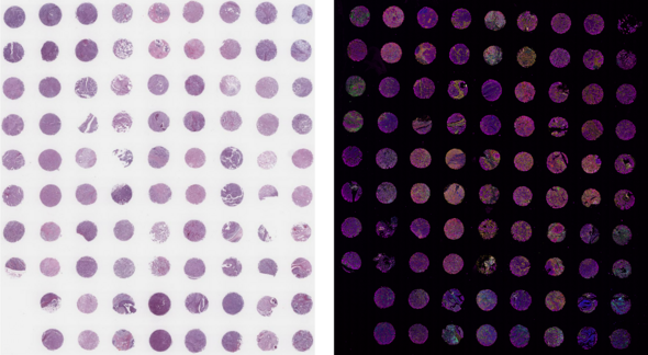
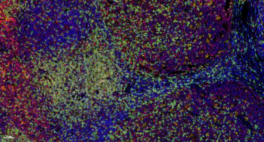
Multiplex immunofluorescence (mIF) is a powerful technique that allows researchers to simultaneously visualize and analyze multiple components of the tissue microenvironment. Multiplexing uses multiple antibodies with fluorescent detection to target specific proteins within the microenvironment, allowing researchers to create a detailed map of the various cells, proteins and their interactions. We offer both low plex and high plex options for mIF which allow you to profile tissues to determine cell phenotypes, their functional state, and cell interactions.

Spatial Biology Workflow

Key Spatial Services
End-to-End Offering
Full Service Histology Lab
Panel Building Expertise
- High- and Low-Plex Options
- In-house Antibody Manufacturing & Validation
- IHC & Carrier-free Antibody Catalog
- Discounted Preset Panels Available
- Fit-for-purpose Panel Development
Our Equipment
PhenoImager™ HT
Lunaphore COMET™

Services
- Up to 6-plex panel using Opal™ reagents
- Panel optimization and antibody order testing
- Library of validated antibodies
- Sample staining
- Slide scanning
Specs
- 0.25 microns/pixel
- Separate 7-colors (DAPI plus 6 antibodies)
- Capable of large tissue microarrays
- Low plex, high throughput, 80 slide capacity
Modular Panel Design
10× Core Panel for human I-O markers
T-cell Profiling Panel
Breast Cancer Module
EMT Panel
Cytokeratin (Structural) Module
Customize Your Panel
| Marker | Host Species | Relevance |
|---|---|---|
| CD3 | Rb | T cells |
| CD20 | Mo | B cells |
| FoxP3 | Rb | Regulatory T cells |
| CD8 | Mo | Cytotoxic T cells |
| PD-L1 | Rb | Checkpoint |
| Pan-CK | Mo | Epithelium |
| PD-1 | Rb | Checkpoint |
| CD68 | Mo | Macrophages |
| CD45 | Rb | Lymphocytes |
| PCNA | Mo | Proliferation |
Our Partners
Quote Request
Spatial Biology CRO Services
Whether
you’re an academic pioneer or a global biotech leader, we deliver comprehensive,
end-to-end spatial biology solutions that prioritize your unique needs, ensuring
you never get lost in the shuffle. Because at Fortis, we believe that your
research deserves more than a one-size-fits-all approach—it deserves a partner
who empowers you.
Multiplex immunofluorescence (mIF) is a powerful technique that allows researchers to simultaneously visualize and analyze multiple components of the tissue microenvironment. Multiplexing uses multiple antibodies with fluorescent detection to target specific proteins within the microenvironment, allowing researchers to create a detailed map of the various cells, proteins and their interactions. We offer both low plex and high plex options for mIF which allow you to profile tissues to determine cell phenotypes, their functional state, and cell interactions.

Spatial Biology Workflow

Key Spatial Services
- High- and Low-Plex Options
- In-house Antibody Manufacturing & Validation
- IHC & Carrier-free Antibody Catalog
- Discounted Preset Panels Available
- Fit-for-purpose Panel Development
End-to-End Offering
- Slide Preparation
- IHC Staining
- Brightfield Slide Scanning
- Multiplex Staining & Imaging
- Image Analysis & Delivery
Full-Service Histology Lab
- Sectioning
- Tissue microarray
- H&E staining
- Whole Slide Scanning
Panel Building Expertise
- Histology services to prepare tissue for staining
- Up to 40-plex discovery panels
- Core panels encompassing basic human I-O phenotypic markers
- Custom validation of antibodies of your choosing
- Library of validated antibodies to choose from
- Creation of custom panels
- Antibodies can be combined with a core panel to create higher plex panels
Our Equipment
PhenoImager™ HT

Services
- Up to 6-plex panel using Opal™ reagents
- Panel optimization and antibody order testing
- Library of validated antibodies
- Sample staining
- Slide scanning
Specs
- 0.25 microns/pixel
- Separate 7-colors (DAPI plus 6 antibodies)
- Capable of large tissue microarrays
- Low plex, high throughput, 80 slide capacity
Lunaphore COMET™

Services
- Runs of 40-plex using seqIF™
- Core Panels available, including SPYRE™ Antibody Panels from Lunaphore
- Addition of other catalog or custom antibodies to Core Panel
- Creation of bespoke panels
- Library of validated antibodies to choose from
Specs
- 0.23 microns/pixel
- 9×9 mm scan area can accommodate tissue sections and small tissue arrays
- No need for conjugated primary antibodies
- High plex, medium throughput
- 24 slides/week for 10-plex
- 20 slides/week for 20-plex
- 8 slides/week for 40-plex
Modular Panel Design
10× Core Panel for human I-O markers
T-cell Profiling Panel
Breast Cancer Module
EMT Panel
Cytokeratin (Structural) Module
| Marker | Host Species | Relevance |
|---|---|---|
| CD3 | Rb | T cells |
| CD20 | Mo | B cells |
| FoxP3 | Rb | Regulatory T cells |
| CD8 | Mo | Cytotoxic T cells |
| PD-L1 | Rb | Checkpoint |
| Pan-CK | Mo | Epithelium |
| PD-1 | Rb | Checkpoint |
| CD68 | Mo | Macrophages |
| CD45 | Rb | Lymphocytes |
| PCNA | Mo | Proliferation |
Extensive collection of in-house manufactured antibodies to choose from to customize your assay
- Immune cell markers
- Cytokeratin markers
- Cancer markers
- Phospho-proteins
- Signaling pathway
- Neurobiology markers
- Mouse proteins


